Neural Circuits and Behavior Research Group
Prof. Dr. Johann Bollmann

Think of taking in the scenery during a hike or moving around in heavy traffic; imagine reading a text or catching a ball. The myriad ways in which we perceive our surroundings and interact with the world critically depend on our eyes, and on how the brain interprets the incoming visual information within fractions of a second. The eyes are our window to the world. To understand how vision works has been at the core of brain research for centuries.
Major building blocks of the human visual system are conserved across all vertebrate species: these include, for example, the lens, which projects an image of our surroundings onto a layer of light-sensitive neurons in the eye’s retina; the retina itself, which is a network of precisely arranged nerve cells in which the image of our visual environment is translated into bioelectrical signals; and likewise, several specialized brain areas that process and interpret these signals coming from the eyes. As a consequence, much of what is known about how vision works has been discovered through research on simpler vertebrate model systems such as fish, amphibians and birds. Yet, vision is far from understood, and therefore the efforts continue: vision labs around the world perform intense research to understand the precise mechanisms that enable us to see, and to understand the causes when vision is impaired or disrupted.
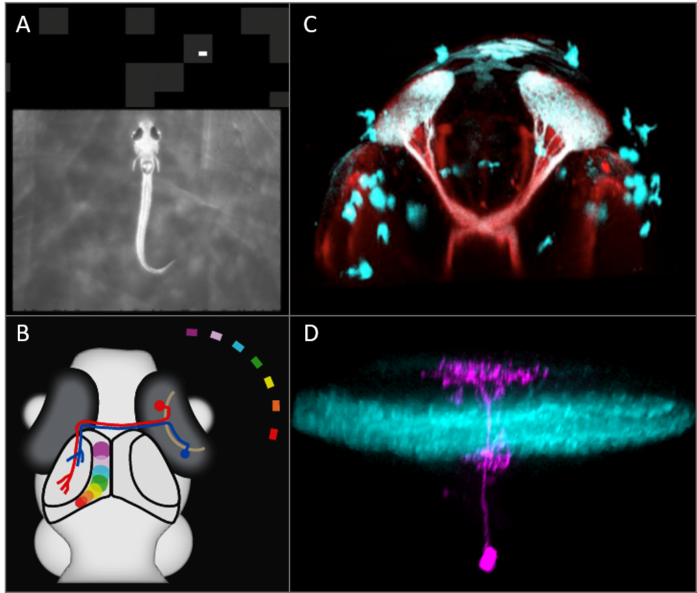
In our research group in Freiburg, we investigate how vision controls action on rapid time scales. We study fundamental neural mechanisms that enable an animal to rapidly analyze its sensory environment, to make behavioral decisions and to generate coordinated motor patterns. We use zebrafish at the larval stage as a neurobiological model as it allows us to investigate the structure and function of the vertebrate visual system and its development in the intact organism. Besides genetic and molecular methods commonly used in the zebrafish model, we use a unique combination of multiphoton calcium imaging, electrophysiological recordings from single, genetically defined cells, and analysis of synaptic connectivity from volume electron microscopy.

